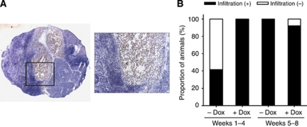
Figure 4.
(A) Effects of wt TP53 induction on metastatic spread of MiaPaCa-2 cells to the liver hilus lymph node. Micro-metastatic spread of MiaPaCa-2-TREx-TP53 cells was evaluated based on detection of human cytokeratin in available mouse lymph nodes (n=7). Inspection of lymph node metastasis, which was stained by pan-cytokeratin antibody. Representative picture of a lymph node stained with an anti-human pan-cytokeratin antibody, revealing positive human tumour cells (stained red) surrounded by lymphocytes (image at × 25 magnification and × 200 magnification (image inset)). (B) Quantitative analysis of lymph node metastasis. Data represent the proportion of animals with presence of lymph node metastasis in the liver hilus lymph node, compared between the treatment (+Dox) and control group (−Dox) of different time periods with wt TP53 reexpression.

