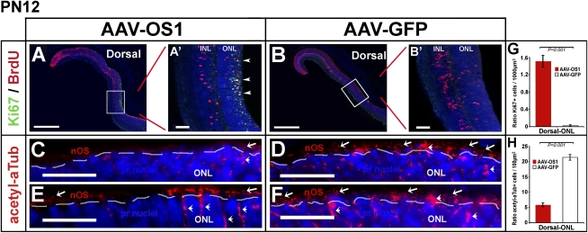
FIGURE 4.
Alteration in the differentiation of photoreceptor cells following Vax2os1 overexpression. (A,B) Fluorescence coimmunostaining for BrdU and Ki67 in the dorsal areas of PN1-injected retinas at PN12. (A′,B′) Higher magnifications of the boxed areas in panels A and B, respectively. There is an increase in the number of the cells positive to both BrdU and Ki67 (arrowheads in A′) in the outer nuclear layer (ONL) of the AAV-OS1-injected retinas in comparison with the AAV-GFP-injected retinal areas. (C–F) Fluorescence immunostaining for acetylated α-tubulin (acetyl-αTub) in the dorsal retinal areas of the injected mice at PN12. There is a decrease in the number of the acetyl-αTub-positive photoreceptor cells (arrows) in the AAV-OS1-injected, as compared with the control-injected retinas. The dotted lines delimit the border between the nOS and the ONL. Note that acetyl-αTub also stains the cytoplasm of Müller glial cells (arrowheads). (G,H) Counts of the Ki67-positive cells (G) and acetyl-aTub-positive photoreceptor outer segments (H) in the dorsal ONL of the injected retinas at PN12. Areas of 160 μm × 40 μm (G) and 80 μm × 5 μm (H) were used for manual counts. For each animal, multiple retinal areas were analyzed. Magnifying bars, 40 μm (A′,B′); 20 μm (C–F). (G,H) Values are means ± SEM (n = 3) and P-values derived from likelihood ratio test for negative binomial.

