
Figure 1.
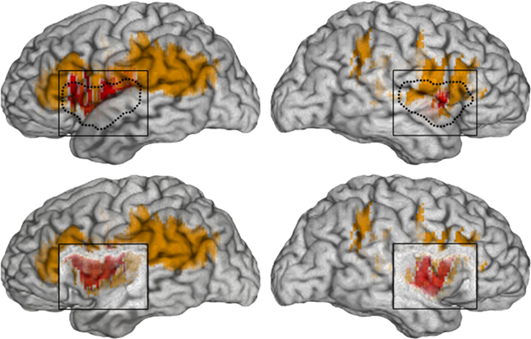
Lesion overlap in the insular cortex lesion group, in views of the right and left lateral surfaces, and with overlaying cortex (insula outlined in dotted lines) in top rows, and with cortex removed exposing the insular cortex underneath in bottom rows. All cases had unilateral lesions in the insular cortex, and the area of damage in the right- or left-sided cases was fairly symmetrical. There is maximal lesion overlap (reflected by red color) across the group in the insular cortex (anterior and posterior) and somatosensory SII region. The lesions in a few patients (reflected by lighter color) were broader and extended posteriorly into the inferior parietal cortex in some subjects, and anteriorly into the inferior frontal gyrus in other subjects.