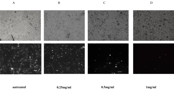
Figure 3.
Fluorescence micrograph of GFP proteins in infected cells treated with DWE. NIH/3T3 cells expressing EGFP following infection with VSV-G pseudotyped HIV-1 virus. (A) NIH/3T3 cells infected with the pLL3.7 lentivirus without DWE (untreated). Figures show NIH/3T3 cells infected with the pLL3.7 lentivirus in the presence of DWE at a concentration of (B) 0.25 mg/ml (C) 0.5 mg/ml (D) 1 mg/ml. In the upper panels, cells are visualized under normal light, while in the lower panels, the same cells are visualized by fluorescent microscope.

