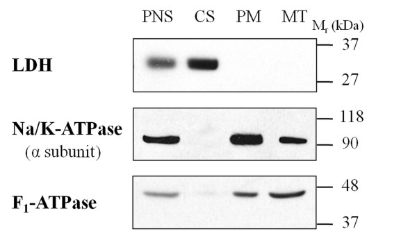
Figure 1.
Distribution of marker proteins in fractionated samples of cardiac tissue. Protein samples (30 μg) of the postnuclear supernatant (PNS), cytosolic (CS), plasma membrane-enriched (PM) and mitochondrial (MT) fractions were subjected to immunoblotting with antibodies against lactate dehydrogenase (LDH), the α subunit of Na, K-ATPase, and F1-ATPase.

