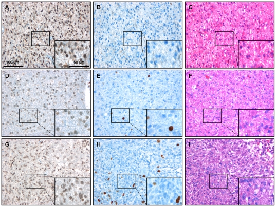
Figure 2. Immunohistochemistry for KLF8 and Ki67 in gliomas of different WHO-grades.
Immunohistochemistry for KLF8 demonstrates that gliomas of different WHO-grades (A: °II, D: °III, G: °IV) show expression of the transcription factor. The expression is ubiquitous in the tissue and shows no grade dependency. Higher magnification demonstrates that KLF8 immunopositive staining is mainly visible in the nuclei but also present in the cytoplasm (Figure 3). Proliferation marker Ki67 is strongly expressed in nuclei of a small population of cells in the same tumors (B: °II, E: °III, H: °IV). Routine HE staining was performed on paraffin embedded sections of all tumor tissue samples (C: °II, F: °III, I: °IV). Scale bars as indicated. For cell count analysis of KLF8 and Ki67 see Table S2.

