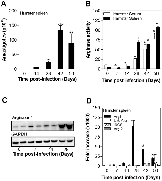
Figure 2. Kinetics of arginase expression in spleen tissue during progressive visceral leishmaniasis. A.
) Parasite burden in the spleens of hamsters (n = 6 per group) infected with L. donovani for 14, 28, 42, and 56 days. The mean and SEM (error bars) of the parasite burden, determined by luminometry and interpolation from an amastigote standard curve, is shown from a single experiment that is representative of 2 independent experiments. B) Arginase activity in the serum (open bars) and spleen tissue (filled bars) of uninfected hamsters (Day 0) and hamsters infected with L. donovani (n = 6 per group) for 7, 14, 28, 42, and 56 days. The mean and SD of the tissue arginase activity, determined by assay of urea production, is shown. C) Hamster arg1 protein expression determined by western blot in spleens of control hamsters (0 days post-infection) and hamsters infected with L. donovani for 7, 14, and 28 days. The expression of GAPDH is shown as a control for protein loading. Each lane contains splenic lysate from a single hamster, with two lanes per time point. The anti-arginase antibody did not react with parasite arginase by immunoblot. D) Time course of expression of hamster arg1 mRNA (filled bars), L. donovani arginase mRNA (empty bars), hamster NOS2 (iNOS) mRNA (hatched bars), and hamster arg2 mRNA (hatched bars) in spleens of control hamsters (0 days post-infection) and hamsters infected with L. donovani for 7, 14, 28, 42, and 56 days. The mean and standard deviation (error bars) of the fold-increase of arginase mRNA relative to BHK cells, determined by real time RT-PCR in groups of 6 animals, is shown from a single experiment that is representative of 3 independent experiments. The statistical significance of differences in each of the panels is identified by asterisks (*, p<0.05; **, p<0.01; ***, p<0.001).

