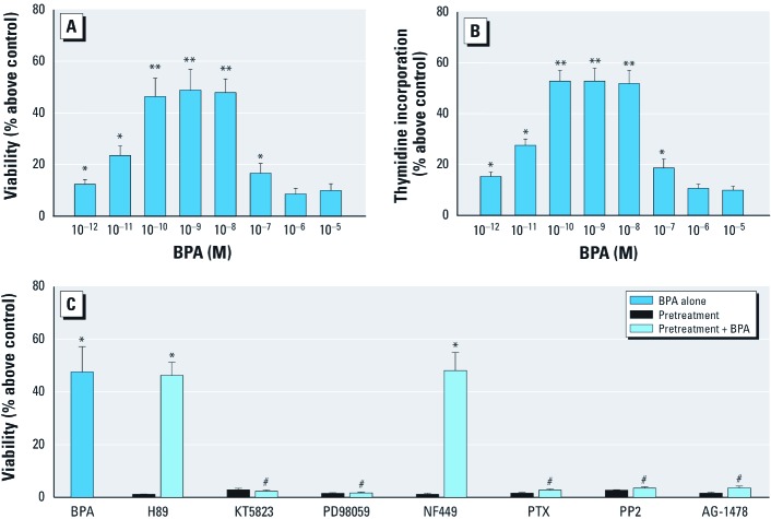
Figure 1.
Twelve-hour exposure to low concentrations of BPA stimulated proliferation of GC-1 cells through activation of the GPCR, PKG, and EFGR-ERK pathways, as determined by the MTT assay (A) and by [3H]-thymidine incorporation (B), as described in “Materials and Methods.” (C) Viability of cells exposed to 10–9 M BPA for 12 hr with or without pretreatment with H89, KT5823, PD98059, NF449, PTX, PP2, or AG1478 (inhibitors of PKA, PKG, MAPK, Gαs, Gαi/ Gαq, Src, and EFGR, respectively). Values shown for A–C are the percentage change above the control (mean ± SE), with the control set as 1; results represent three independent experiments performed in triplicate. *p < 0.05, compared with control. **p < 0.05, compared with 10–12, 10–11, 10–7, 10–6, or 10–5 M BPA. #p < 0.05, compared with 10–9 M BPA alone.

