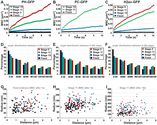
Figure 6. The fast local motion of PC bodies is less constrained than that of chromatin domains.
A–C: Mean square change (MSC) monitoring the motion of PC bodies (PH-GFP: A and PC-GFP: B) and chromatin domains (H2Av-GFP: C) during embryogenesis. The kinetics of PC bodies monitored by PC-GFP or PH-GFP are similar throughout embryogenesis. Although PC bodies and chromatin domains similarly slow down during development, chromatin domains consistently move less than PC bodies (p<0.001, KS test calculated with MSC values reached after 1 s). D–F: Histograms presenting the frequency of angles calculated between three consecutive time-points in tracks of PC bodies (PH-GFP: D and PC-GFP: E) or chromatin domains (F). Narrow angles are over-represented in tracks of PC bodies and condensed chromatin domains throughout embryogenesis. G–I: Scatter plots comparing the average radius of the volume in which PC bodies (PH-GFP: blue points and PC-GFP: red points) or chromatin domains (black points) move, with the distance between the two objects tracked.

