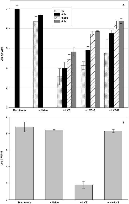
Figure 3. Splenocytes from mice vaccinated with a panel of LVS-related vaccines exhibit a hierarchy of control of intramacrophage LVS growth.
(A) BMMØs from C57BL/6J mice were infected with LVS at an MOI of 1∶20 (bacterium-to-macrophage ratio; “Mac alone”), and co-cultured with the indicated numbers of splenocytes obtained from either naive C57BL/6J mice or C57BL/6J mice vaccinated 1×10⁴ CFU LVS, 1×10⁴ LVS-G, or 1×10⁴ LVS-R 6 weeks previously. Here, for all co-cultures containing added splenocytes, “1x” = 5×10⁶ splenocytes per well (used in all previous experiments), and 0.5x, 0.25x, and 0.1x refer to corresponding decreases in the total number of added splenocytes. (B) BMMØs from C57BL/6J mice were infected with LVS at an MOI of 1∶20 (bacterium-to-macrophage ratio), and co-cultured with splenocytes obtained from either naive C57BL/6J mice or C57BL/6J mice vaccinated 1×10⁴ CFU LVS or 1×10⁸ HK-LVS 6 weeks previously. For both A and B, after three days of co-culture, BMMØ were washed, lysed, and plated to determine the recovery of intracellular bacteria. Values shown are the mean numbers of CFU/ml ± SD of viable bacteria for triplicate samples. Results shown are from one representative experiment of three (A) or four (B) independent experiments of similar design with similar outcome.

