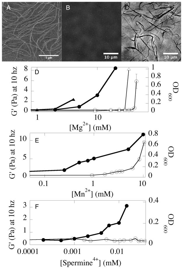
FIG. 1.
Gelation and bundling of Pf1 virus by divalent and tetravalent counterions. An image of single Pf1 viruses in monovalent salt taken by atomic force microscopy (A). The same sample of Pf1 (0.01 % w/v) looks transparent under phase contrast light microscopy (B) but bundles of Pf1 form after addition of 50 mM MgCl2 (C). Storage shear modulus (G′ measured at 2% strain and 10 rad/s; closed symbols) and optical density (open symbols) of 0.5% Pf1 in solutions containing 2 mM HEPES, pH 7.5 and various concentrations of Mg2+ (D) Mn2+ (E) or sperimine4+ (F). Gelation and aggregation of a modified Pf1 with increased surface charge (triangles) are compared to those of normal Pf1(circles) in (D).

