
Figure 2. Morphology and magnetism of collagen-SPION nanobiocomposite.
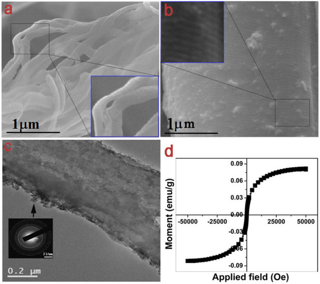
FESEM showing (a) collagen waste fibers (b) collagen waste-SPION nanobiocomposite fiber bundle. Insets show the magnified view of the select portion exhibiting the bands of collagen fibers; (c) TEM of collagen waste-SPION nanobiocomposite fiber showing the SPION coated on the collagen fiber. SAED shows the crystalline nature of the SPION; (d) SQUID of collagen waste-SPION nanobiocomposite showing superparamagnetism.