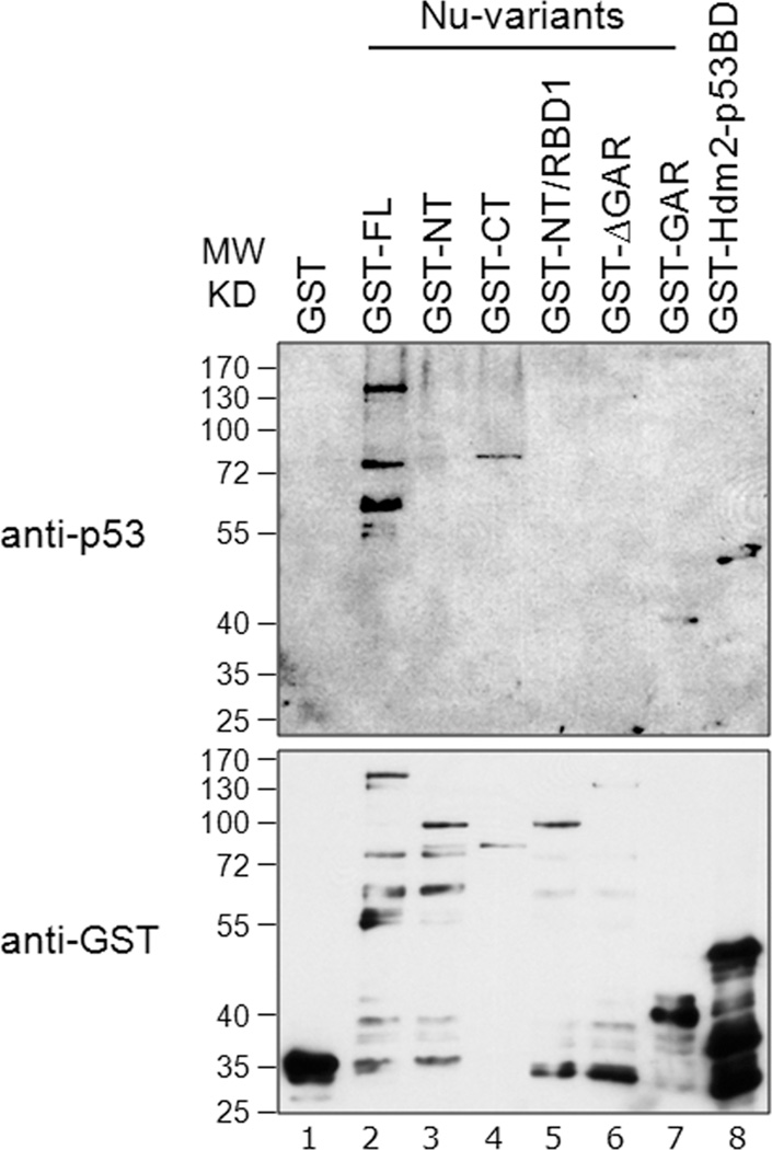
Fig. 5. Nucleolin GAR interacts with p53.
Equivalent amounts (200 ng) of GST-tagged nucleolin-FL (lane 2) or truncation domains (lanes 3 to 7) were subjected to SDS-PAGE. After transfer to a nitrocellulose membrane, the membrane was probed with purified GST-p53 protein (0.2 µg/ml) in a Far-Western assay (upper panel, lanes 1 to 8). The bound p53 was then visualized using a monoclonal anti-p53 antibody (DO-1). To visualize GST-tagged proteins, a parallel blot was run and probed directly for anti-GST antibodies (lower panel, lanes 1 to 8). GST alone (lane 1) and GST-tagged Hdm2 p53-BD (lane 8) were used as negative and positive binding controls, respectively.

