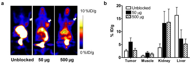
Fig. 4.
a Decay-corrected whole-body coronal microPET images of UM-SCC1 tumor–bearing mice (right shoulder) at 30 min after injection of 3.7 MBq (100 μCi) of [18F]FBEM-cEGF without or with 50 and 500 μg of unlabeled hEGF. Tumors are indicated by white arrows. b Quantification of [18F]FBEM-cEGF uptake in UM-SCC1 tumors, muscle, liver, and kidney. Data are presented as mean %ID/g ± SD (n=3).

