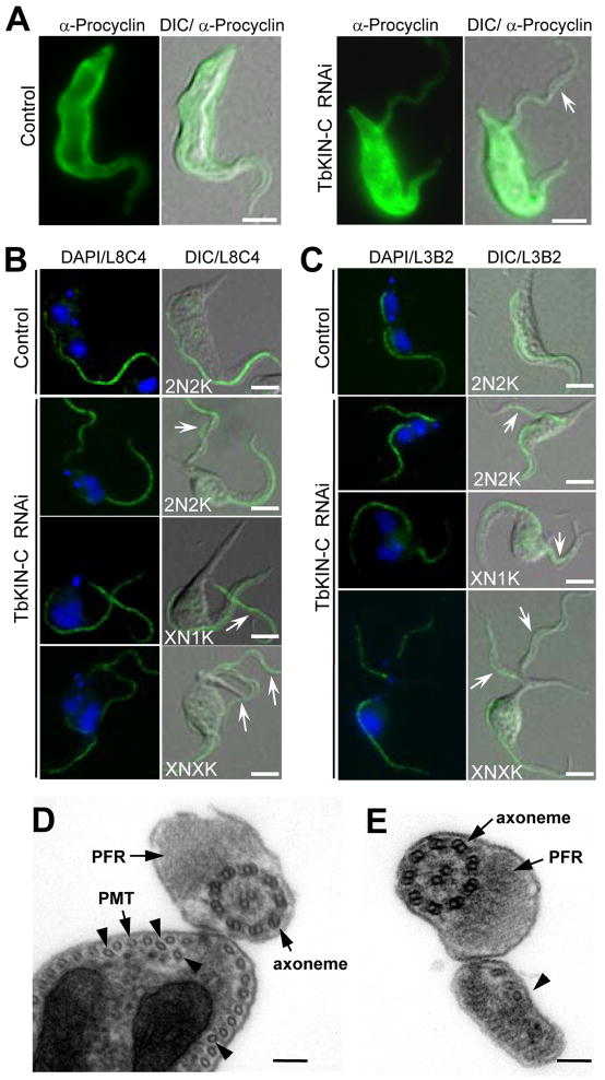
Figure 4. Effect of TbKIN-C RNAi on the synthesis of flagellum and flagellum attachment zone.
(A). Immunostaining of control and TbKIN-C RNAi cells with anti-Procyclin antibody. The arrow indicates the detached flagellum that contains the membrane and cytoplasm. Bars: 5 μm. (B). Effect of TbKIN-C RNAi on the growth of the new flagellum. Cells were fixed in cold methanol, stained with L8C4 antibody for flagella, and counterstained with DAPI for DNA. Arrows point to the detached flagella. Bars: 5 μm. (C). Effect of TbKIN-C RNAi on growth of the new flagellum attachment zone. Cells were fixed in cold methanol, stained with L3B2 antibody for FAZ, and counterstained with DAPI for DNA. Arrows point to the detached flagellum that contains full-length FAZ. Bars: 5 μm. (D, E). Cross-section through the attached flagellum (D) and the detached flagellum (E) of TbKIN-C RNAi cells. The flagellar axoneme, paraflaegllar rod (PFR), and the subpellicular microtubules (PMT) are indicated. The arrowheads in panel D indicate the disorganized PMT, and the arrowhead in panel E points to the portion of cytoplasm detached together with the flagellum. Bars: 100 nm.

