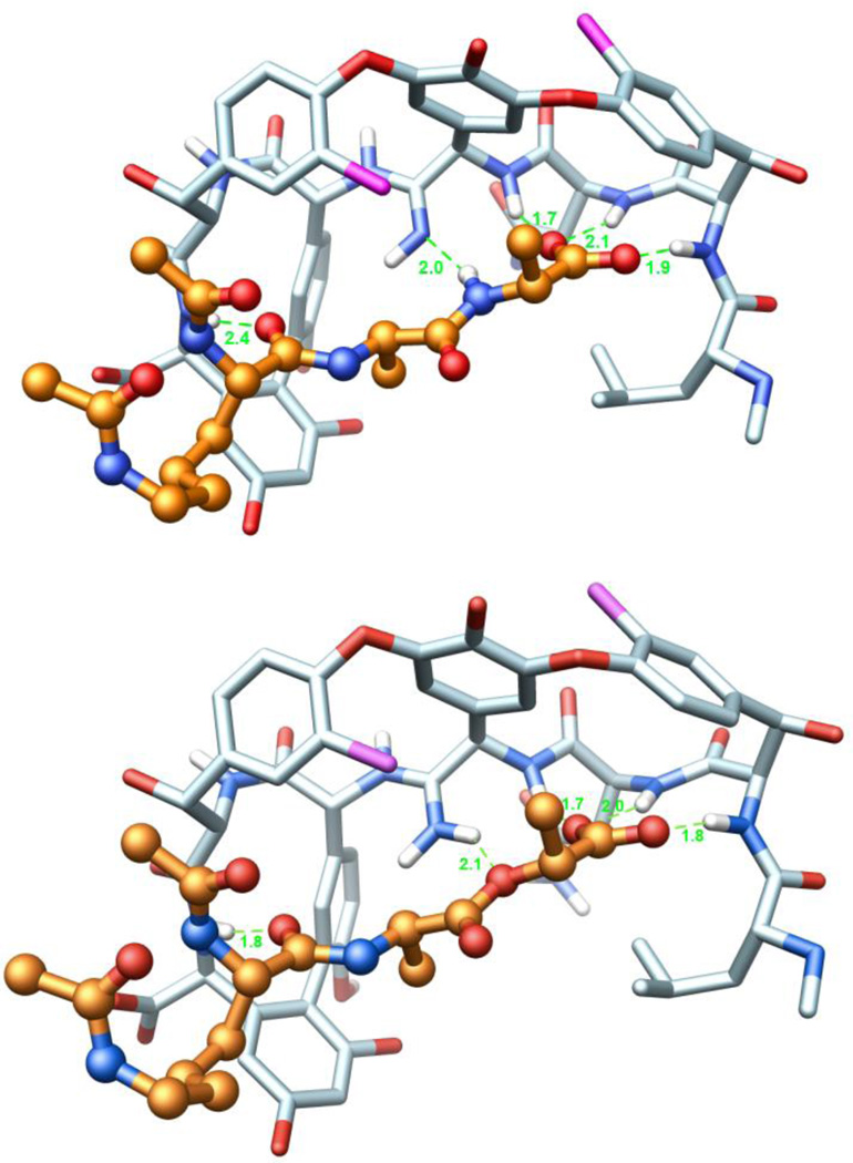
Figure 9.
(Top) Ball-and-stick representation of a energy minimized structure (MOLOC34) of 7 (C-atoms: light blue) and (Ac)2-l-Lys-d-Ala-d-Ala (C-atoms: salmon). (Bottom) Ball-and-stick representation of a energy minimized structure of 7 (C-atoms: light blue) and (Ac)2-l-Lys-d-Ala-d-Lac (C-atoms: salmon). Hydrogen atoms of all amides are depicted in white, H-bonds are depicted as green-dotted lines, and H-bonding distances are indicated. The corresponding H-bond distances for vancomycin binding to (Ac)2-l-Lys-d-Ala-d-Ala (see Figure 5) in the X-ray structure are 1.9, 1.9, 1.8, 2.0, and 1.7 Å, respectively. Structures obtained by modification of a co-crystal structure of vancomycin aglycon and (Ac)2-l-Lys-d-Ala-d-Ala (PDB code: 1FVM).

