Fig. 7.
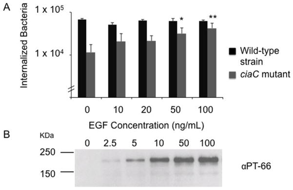
Stimulation of the EGF signaling pathway rescues the invasiveness of a C. jejuni ciaC mutant. Panels: A) INT 407 cells were incubated with the concentrations of EGF indicated for 30 minutes prior to inoculation with either the C. jejuni wild-type strain or the ciaC mutant. Values represent mean ± standard deviation of internalized bacteria/well of a 24-well tissue culture tray. The asterisks (* P < 0.05, ** P < 0.01) indicate a significant increase in the number of internalized bacteria for cells treated with EGF and inoculated with the ciaC mutant versus untreated cells inoculated with the ciaC mutant. B) Immunoblot probed with the PT-66 α-phosphotyrosine antibody. INT 407 cells were treated with EGF and cell lysates prepared after a 30 min incubation period. The blot was exposed for a short period of time in order to show that the EGFR (Mr = 180 kDa) is activated in a dose-dependent manner in response to increasing concentrations of EGF.

