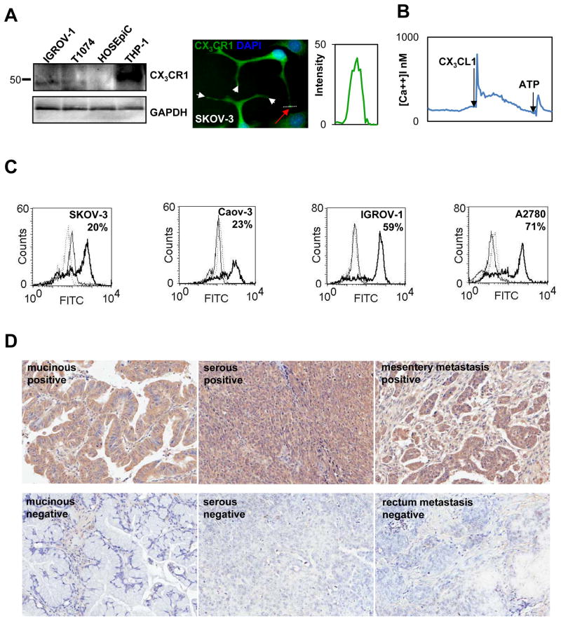
Figure 3. CX3CR1 is expressed in primary and metastatic epithelial ovarian carcinoma.
(A) CX3CR1 expression in normal ovarian surface epithelial cells (HOSEpiC), immortalized normal ovarian surface epithelial cells (T1074), and serous EOC cell line (IGROV-1) was evaluated by Western blot. GAPDH served as a loading control. THP-1 cell lysate was used as a positive control for CX3CR1 expression. Position of the marker lane of apparent molecular weight of 50 kDa is indicated. Expression of CX3CR1 in the serous EOC cell line (SKOV-3) was evaluated using immunofluorescence staining. CX3CR1 – green; DAPI – blue. Images were collected independently on the green and blue filters and subsequently superimposed. Bar, 10 μm. Histogram shows the intensity of CX3CR1 staining across the pseudopodial protrusions of SKOV-3 cells (white dotted line and red arrow). (B) Intracellular [Ca++] changes in response to 20 nM CX3CL1 indicate the presence of active CX3CR1 in EOC cells. An ATP-induced signal indicates live cells. (C) Cell surface expression of CX3CR1 in the indicated serous EOC cell lines was analyzed by flow cytometry. The percentage of positive cells is indicated. Solid thick line – CX3CR1 expression; solid thin line – negative control (normal rabbit IgG and FITC-conjugated goat anti-rabbit IgG); dotted line – negative control (FITC-conjugated goat anti-rabbit IgG only). These data are representative of at least three independent experiments. (D) Expression of CX3CR1 in cases of primary and metastatic EOC was determined by immunohistochemistry. Brown – CX3CR1; blue – hematoxylin. Images were generated using an Aperio ScanScope digital slide scanner. Magnification - 10×. Examples of CX3CR1-positive and CX3CR1-negative cases of mucinous adenocarcinoma (cores C10 and C5, respectively, Supplemental Table 1), serous carcinoma (cores I15 and G9, respectively, Supplemental Table 1), and metastatic serous carcinoma (cores J8 and L3, respectively, Supplemental Table 1) are displayed.

