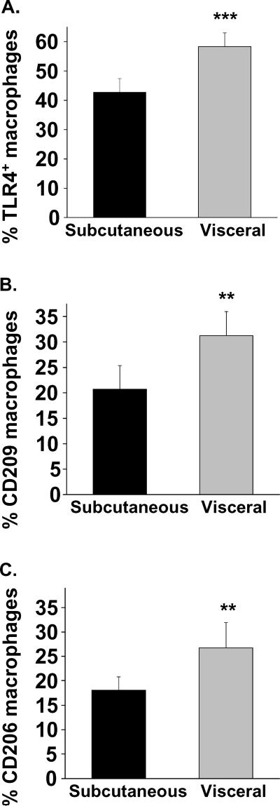
Figure 3. Depot-specific macrophage polarization in adipose tissue.
Adipose flow cytometry identified higher populations of (A) TLR4+ M1 polarized macrophages in visceral vs. sc adipose tissue. Similarly, macrophages expressing markers traditionally associated with M2 phenotypes (B) CD209 and (C) CD206 were also significantly higher in visceral fat. ***p<0.001; **p<0.01 compared to subcutaneous depot. Data presented as mean ± SEM.

