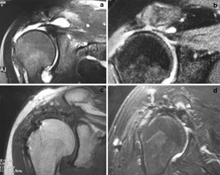
Fig. 1.
Preoperative coronal T2-weighted magnetic resonance image (MRI) showing complete injury with a 3-cm retraction in 2 patients (a, b). Coronal T1-weighted (c) and T2-weighted (d) MRI sequences showing postoperative findings with tendons repaired, blooming artifacts at footprint, high-intensity tissue at the footprint (c), and high-intensity tissue at tendon–bursa interface (d)

