
Fig. 4. Mutational analysis of the putative ligand-binding cavity of AphB.
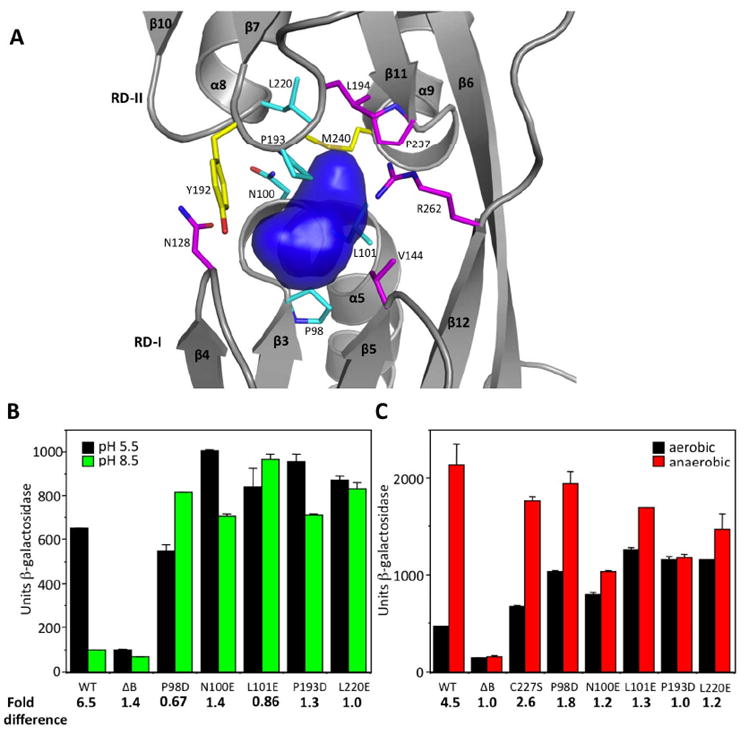
(A) Surface representation of the putative ligand-binding cavity of AphB (blue) generated with SURFNET (Laskowski, 1995). Residues lining the cavity were substituted with negatively-charged amino acids. Most of these substitutions resulted in loss (magenta) or gain (cyan) of function, whereas two (yellow) did not affect activity. (B) Influence of the ligand-binding pocket substitutions (P98D, N100E, L101E, P193D, L220E) on the expression of the tcpPH promoter in V. cholerae at pH 5.5 (black bars) and pH 8.5 (green bars). (C) The same ligand-binding pocket substitutions as in (B) and C227S on the expression of the tcpPH promoter in V. cholerae at neutral pH under aerobic (black bars) and anaerobic conditions (red bars).