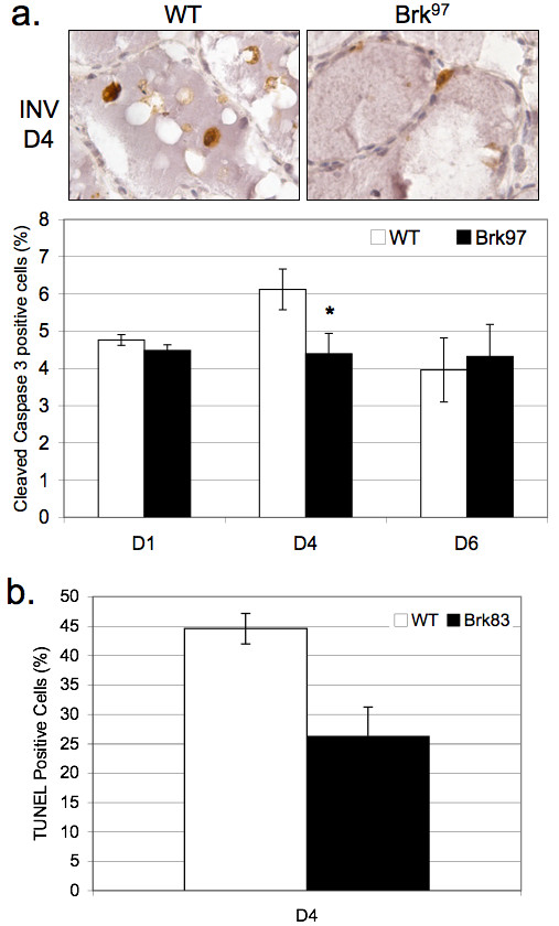
Figure 3.

Apoptotic events are decreased in mammary tissue from WAP-Brk mice. A, Cleaved caspase 3 IHC. Images: INV D4 in wild-type and Brk97 mice, 400 × magnification. Graph: Quantification of positive IHC staining, presented as a percentage of total mammary epithelium. B, Quantification of Terminal deoxynucleotidyl transferase mediated dUTP Nick End Labeling (TUNEL) positive cells in INV D4 mammary gland sections.
