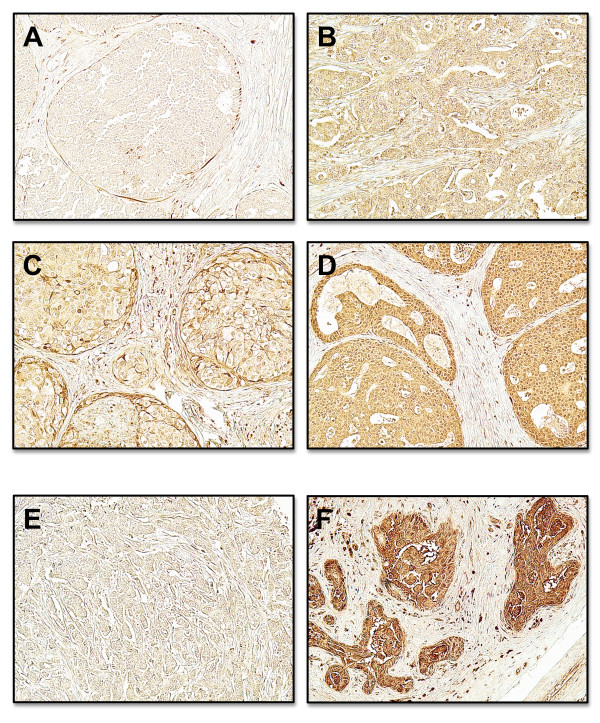
Figure 1.
Expression of CX3CR1 in human breast cancer tissue arrays. Panel A shows a representative sample that stained negative for CX3CR1. The majority of samples examined showed different degrees of positive staining for the receptor in the epithelial cells (B-D; see also Table 1). Panels E and F show a negative and highly positive sample for CX3CR1, respectively, at higher magnification. The stromal compartment stained uniformly negative for CX3CR1. Representative images of 47 normal and 202 malignant tissue cores analyzed. (Original magnification ×100 for A to D and ×200 for E and F).

