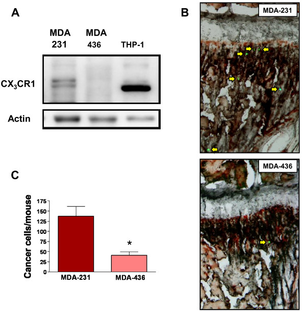
Figure 2.
Expression of CX3CR1 by human breast cancer cells. Western blotting analysis of CX3CR1 expression in MDA-231 and MDA-436 breast cancer cell lines. CX3CR1 was detected in MDA-231 cells, whereas MDA-436 showed negligible levels of the receptor. The two bands observed are likely due to the detection of different isoforms of the receptor produced by alternative splicing [33]. Cell lysates from the THP-1 human monocytic cell line were included as a positive control. Actin was used as a loading control (A); single breast cancer cells stably expressing eGFP (arrows) were identified by fluorescence stereomicroscopy in the bone marrow of mice, 24 hours after being inoculated via the left cardiac ventricle (B); MDA-231 and MDA-436 cell lines that migrated to the femora and tibiae of mice inoculated via the hematogenous route, expressed as mean of cells detected per animal + S.E.M. (C). (MDA-231 cells = four mice, MDA-436 cells = eight mice. * P = 0.0008).

