Figure 7.
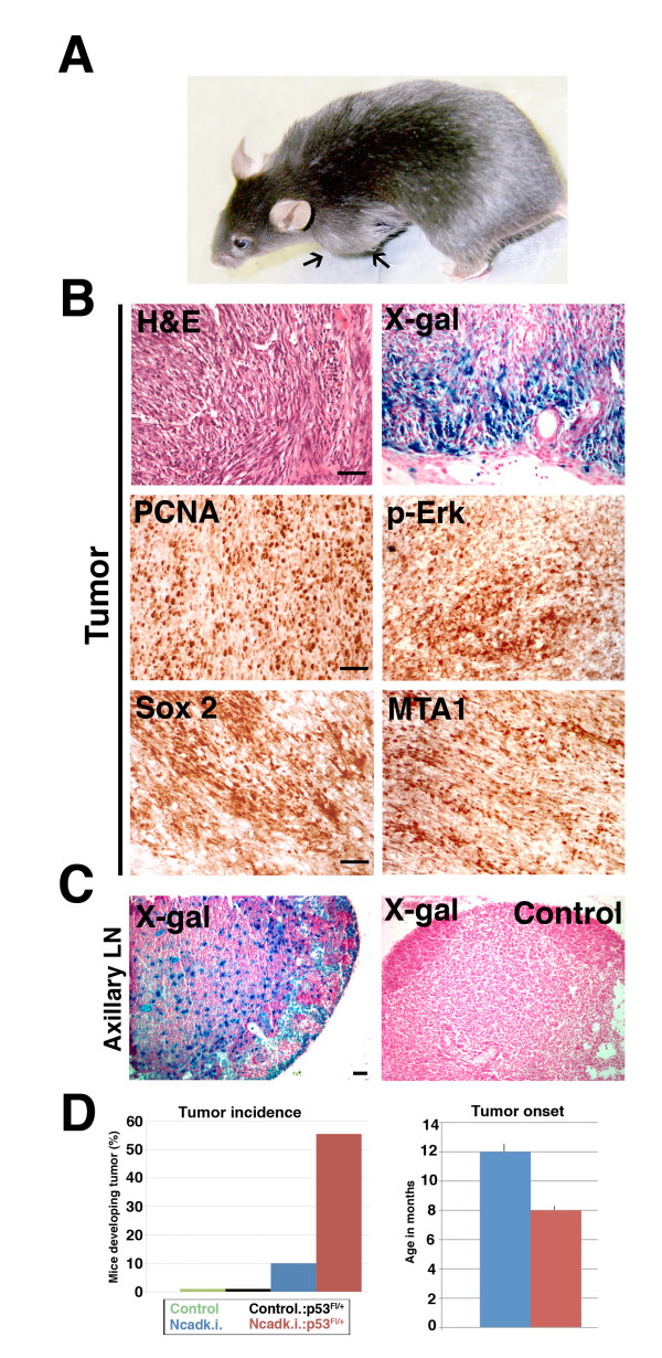
Ncadk.i. and Ncadk.i.;p53 females develop malignant and highly invasive tumors. (A) Ncadk.i. and Ncadk.i.;p53 females developed large tumors (arrows) after numerous lactation cycles that were always found in the left thoracic mammary gland. (B) H&E staining of tumor sections revealed poorly differentiated cells. X-gal staining identified many transformed alveolar epithelial cells, which contribute to the tumor structure. Immunohistochemistry on paraffin sections revealed that tumor cells are highly proliferative, indicated by PCNA staining, and express high levels of p-Erk, Sox2, and the metastatic marker MTA1. Scale bar: 100 μm (C) X-gal staining traced alveolar epithelial cells derived from the tumor in the axillary lymph node, demonstrating the metastatic capacity of the tumor cells. No LacZ-positive cells were found in control lymph nodes. Scale bar: 100 μm. (D) Heterozygous deletion of p53 enhances the tumorigenic potential of N-cad. At least nine animals were analyzed for tumor incidence and tumor onset. Ten percent of Ncadk.i. mice develop mammary tumors. This number increases to more than 50% upon additional heterozygous deletion of p53 (left panel). The average age of tumor onset is decreased in Ncadk.i.;p53 compared to Ncadk.i. mice (right panel). H&E, Hematoxylin and Eosin; MTA1, Metastatic tumor antigen1; Ncadk.i., WAP::Cre;EcadNcad/fl; Ncadk.i.;p53, WAP::Cre; EcadNcad/fl;p53fl/+; p53, protein 53; p-Erk, phospho-extracellular signal-regulated kinase; Sox2, SRY (sex determining region Y)-box 2; X-gal, 5-bromo-4-chloro-indolyl-galactopyranoside.

