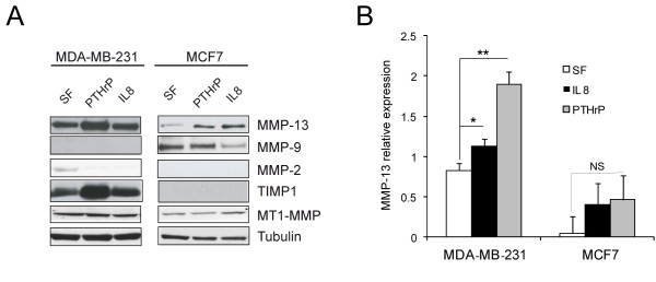
Figure 2.
MMP expression in MDA-MB-231 and MCF7 cells. A. MMP expression in MDA-MB-231 and MCF7 cells, stimulated with PTHrP or IL-8 for 24 h in serum free medium. Protein extracts of cellular lysates (for detection of MT1-MMP) and supernatants (for detection of MMP-13, MMP-9, MMP-2 and TIMP-1) were loaded for Western blotting analysis. Representative blottings are reported. Tubulin was used as a loading control. B. Quantification of Western blot analysis for MMP-13 expression by QuantityOne software (Bio-Rad; Milan, Italy). Mean values (± SD) of MMP-13 relative expression levels of three different experiments are reported. *P < 0.05; **P < 0.01; NS, not significant.

