Figure 6.
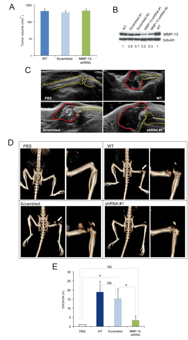
MMP-13 increases osteoclastogenic potential in vivo. WT and transfected cells were inoculated into the femurs of 6 weeks-old nude mice. Volume (A) and MMP-13 expression (B) of the tumours developed one month after the inoculation of WT, scrambled and shRNA MDA-MB-231 cell clones. Data in (A) are expressed as the mean ± SE (WT, n = 8; Scrambled, n = 11; MMP-13-shRNA, n = 12). C. Representative ultrasound images of WT, scrambled and shRNA #1 clones after one month from injection into nude mice. The basal skeletal condition is represented by PBS inoculation. Red and yellow dotted lines approximately label tumour mass and bone profile, respectively. D. Representative images and their magnification of CT analysis of skeletal lesions produced by MDA-MB-231 cell clones injected into nude mice. E. The graph shows the level of femur erosion, calculated as follows: (1- (length of the undamaged femur injected/length of untreated counterpart)) × 100. Results are the mean ± SE (WT, n = 8; Scrambled, n = 11; MMP-13-shRNA, n = 12). *P < 0.05; NS, not significant.

