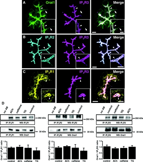
Figure 1. Orai1 co-localization and co-immunoprecipitation with IP3Rs in PACs.
(A) Maximum projection of 20 optical sections spaced 1 μm from each other in a PAC cluster. Orai1 (green) is present in both the basolateral (arrows) and apical (arrowheads) regions. In the apical pole, Orai1 co-localizes with IP3R3 (magenta). Insets: single confocal sections from the same cluster at higher magnification (the arrow and arrowhead points to the same structures as in the main Figure). Here and in (B) and (C) the scale bars correspond to 10 μm in the projections and 5 μm in the insets. (B) Maximum projection of optical sections of a PAC cluster. IP3R2 (cyan) co-localizes with IP3R3 (magenta) in the apical pole of the cells (arrowhead). Insets: single confocal sections from the same cluster at higher magnification (the arrowhead points to the same structures as in the main Figure). (C) Maximum projection of optical sections of a PAC cluster. The highest density of IP3R1 (yellow) is observed in the apical pole of the cells (arrowheads), where it co-localizes with IP3R3 (magenta). Insets: single confocal sections from the same cluster at higher magnification (the arrowhead points to the same structures as in the main Figure). Note that a significant staining for IP3R1 (unlike that for IP3R2 and IP3R3) was also found outside the apical region of the cell. (D) Co-immunoprecipitation of Orai1 with IP3R1 (left panel), IP3R2 (middle panel) or with IP3R3 (right panel) in PAC lysates. The first lane in both panels corresponds to beads that were not bound to anti-IP3R antibodies. Western blots show the IP3Rs and Orai1 eluted from Sepharose beads decorated with the corresponding IP3Rs. Histograms show the quantification of Western blots.

