Figure 16.
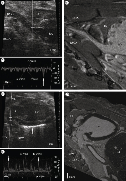
Micro-ultrasound imaging of the right and left atrial inflow channels, with anatomical confirmation by magnetic resonance (MR) imaging. (a) Image of a right parasternal longitudinal section. (b) The Doppler flow spectrum of right superior vena cava (RSVC), showing a small retrograde wave caused by atrial contraction. (c) MR image of a similar section to the micro-ultrasound image in (a), showing the vascular continuity from RSVC to the right atrium (RA), and the surrounding organs such as thymus (TH), right subclavian artery (RSCA), right pulmonary artery (RPA) and right pulmonary vein (RPV). (d) The micro-ultrasound image of a left parasternal longitudinal section showing the RPV, LA and LV, with the Doppler sample volume in the entrance of the RPV. (e) The Doppler flow spectrum from PV. The arrows indicate heart beats at the end of inspiration. (f) The MR image of a similar section to the micro-ultrasound image in (d). Adapted from [22].

