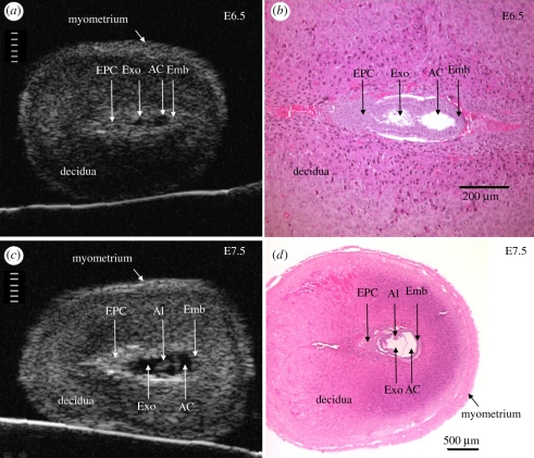
Figure 3.
Anatomical detail visible in ultrasound images of the conceptus in the exteriorized uterus at E6.5 and E7.5. Ultrasound images (a,c) and H and E histological sections (b,d) of implantation sites at E6.5 (a,b) and E7.5 (c,d). Divisions in the scales in (a) and (c) are 100 µm apart. The conceptus in histological sections is smaller than in vivo due to shrinkage during tissue preparation (fixation and dehydration). AC, amniotic cavity; Al, allantois; Emb, embryo; EPC, ectoplacental cone region; Exo, exocoelomic cavity. Adapted from [48].

