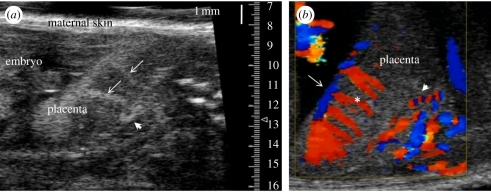
Figure 7.
Micro-ultrasound imaging of the placental circulation in the mouse. (a) Contrast enhanced visualization of the uteroplacental blood supply to the mouse placenta using MBs infused into the maternal circulation at E14.5. A spiral artery in the decidua is indicated by the arrowhead. Arrows show the maternal arterial canals. Canals are formed by foetal trophoblast cells and they direct maternal blood from the spiral arteries into the labyrinth, the exchange region of the placenta. (b) Colour Doppler imaging of the uteroplacental and foetoplacental blood supply to the mouse placenta at E14.5. A spiral artery is shown by the arrowhead. The spiral movement of blood in the maternal spiral artery causes the red–blue alternating pattern as the blood alternates between flowing towards and away from the transducer. Flow in the foetal chorionic plate vessels (arrow) and in the foetoplacental arterioles (asterisk) directs foetal blood deep into the labyrinth exchange region of the placenta.

