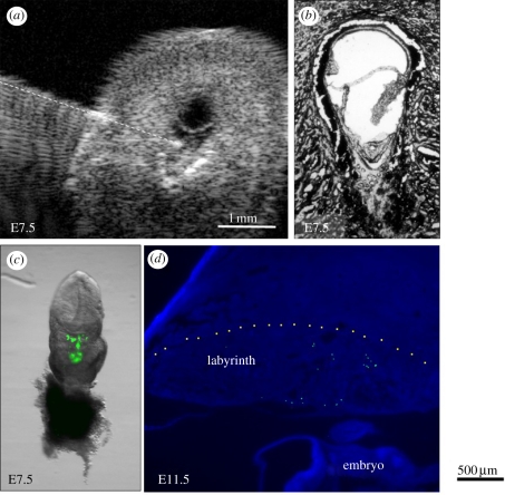
Figure 9.
Ultrasound-guided microinjection of green fluorescent microspheres into the exocoelomic cavity of a mouse conceptus at E7.5. (a) The glass microinjection catheter (highlighted with a dashed line) is shown with its tip in the exocoelomic cavity in an exteriorized conceptus. (b) Histological image showing the anatomy of the conceptus at E7.5. (c) A conceptus dissected on the day of injection showing green fluorescent microspheres in the exocoelomic cavity. The embryo will form above and the placenta below this cavity. (d) A placenta dissected later in gestation at E11.5 showing green fluorescent microspheres confined within the labyrinth region of the placenta. Adapted from [48].

