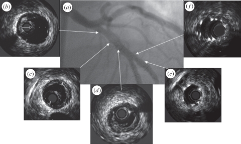
Figure 2.
(a) Angiogram with corresponding IVUS echograms (b–f) of a coronary artery demonstrating that the luminal area as provided by the echogram does not provide information on the presence of plaque (b,c). Furthermore, the presence of (d) a dissection and (e) the adequate positioning and (f) malapposition of a stent can be visualized by IVUS.

