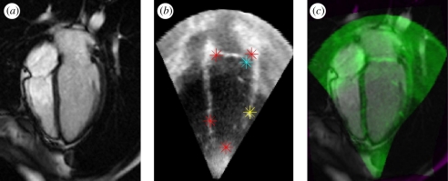
Figure 4.
Two-dimensional non-rigid alignment of echocardiography and cardiac MRI. (a) Cardiac MRI image, (b) ultrasound image with red, blue and yellow markers indicating points about which local alignment corrections are made at successive iterations of the registration algorithm. (c) The green overlay is the echocardiography image superimposed on the cardiac MR image slice. (Result courtesy of Weiwei Zhang, U. Oxford, UK).

