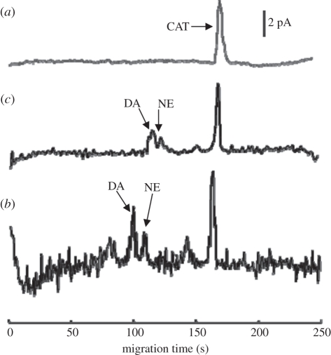
Figure 10.
Electropherograms for three different media as follows: (a) catechol in HEPES buffer as the standard indictor (5 nM), (b) culture medium from living PC-12 cells after nicotine stimulation (two to three cells), and (c) culture medium from PC-12 cells after cell lysis by a high-concentration KCl solution.

