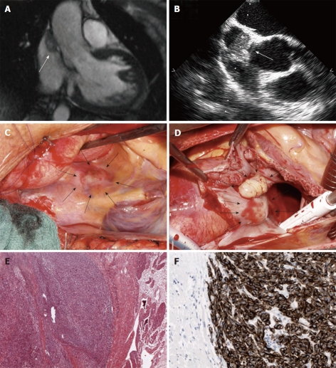
Figure 2.
Tumor imaging and histology. Magnetic resonance imaging (A) and transesophageal echocardiography (B) showing the tumor in the inflow of the right atrium. Intraoperative views of the superior vena cava from the outside (C) and the inside (D); Histologic examination reveals intracardiac metastasis of a malignant melanoma (E, F). Arrows indicate tumor localization.

