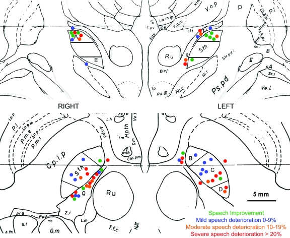
Figure 1. Location of the active contacts at 1 year post bilateral subthalamic nucleus deep brain stimulation (STN-DBS).
Location of the active contacts in 31 patients at 1 year post bilateral STN-DBS as transposed onto the Schaltenbrand atlas adopting the radiologic imaging convention (right STN on the left side of the image). Top: coronal view adapted from plate 27, f.p. 3.0. Contacts related to the superior (A) and inferior (E) segment of the STN are shown. The middle section of the STN in coronal view is further subdivided into 3 segments in the axial plane shown below. Therefore, the coronal and axial views show different contacts. Bottom: axial view adapted from plate 55, H.v. 4.5. Contact location is shown in relation to the anteromedial (B), central (C), and posterolateral (D) segments of the STN. Selected abbreviations: Ru = red nucleus; Sth = subthalamic nucleus; Z.i. = zona incerta. (Scaltenbrand G, Wahren W. Atlas for Stereotaxy of the Human Brain, 2nd ed. New York: Thieme; 1977. Plates 27;55. Reprinted by permission.)

