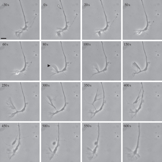
Figure 1.
Directed filopodial extension in response to a laser injury 20 µm behind a growth cone. Following irradiation at the site indicated by the arrow (0 s), thinning of the axon was observed beginning at 20 s followed by restoration of thickness by 150 s. At 80 s, a large filopodium indicated by the arrowhead has formed nearest the lesion site. By 300–350 s, it has extended and contacted the axonal shaft near the lesion site. The filopodium regresses forming a large vesicle at 450–550 s. At 600 s, the growth cone resumes growing in the pre-irradiation direction. Scale bar, 5 µm.

