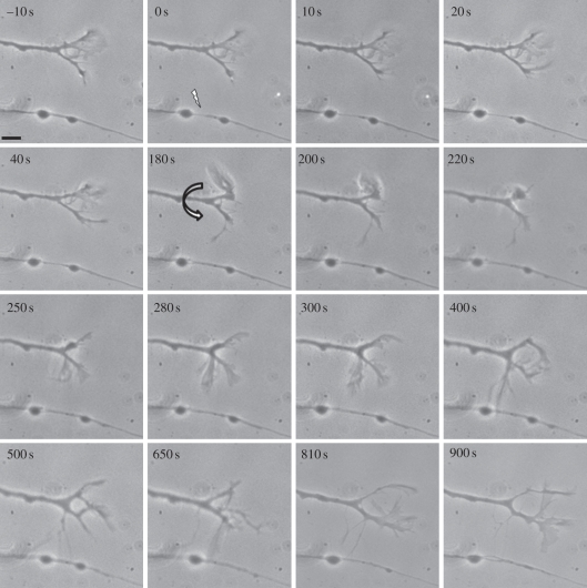
Figure 2.
Directed filopodial extension in response to laser injury on a parallel growing axon. Irradiation site is indicated by the arrow. Irradiation at 0 s leads to thinning of the axon shaft. At 180 s, the non-irradiated growth cone extends a filopodium towards the injury site, makes apparent contact at 300–400 s, and subsequently withdraws. The growth cone continues moving in its original direction. Scale bar, 5 µm.

