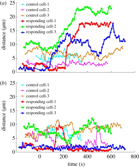
Figure 4.
Plots of the longest filopodium extending (a) towards the damaged axon or (b) away from the axon as a function of time. (a) Filopodia were measured in the ‘toward zone’ extending from the growth cone and subtending a 10 µm region centred along the injured axon. (b) As a comparison, filopodia were measured in the mirror image of the ‘toward zone’ triangle reflected across the growth cone in the ‘away zone’. (Online version in colour.)

