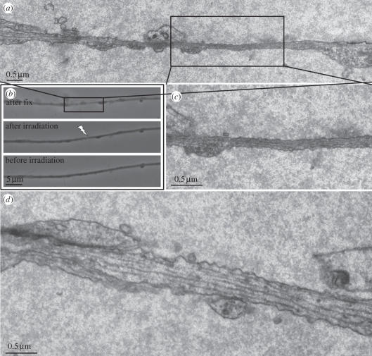
Figure 8.
TEM and phase contrast images of damaged axon: (a) reconstructed collage of multiple TEM images of an axon fixed 30 s after laser irradiation. (b) Live phase contrast images taken before (bottom) and after irradiation (middle) and after fixation (top). Images are matched with the electron microscope images in (a). (c) Electron micrograph of the region in the centre of the ‘thinned’ zone. Note the intact cell membrane and the presence of contiguous microtubules. (d) Non-irradiated region 36 µm away from the laser-irradiated region.

