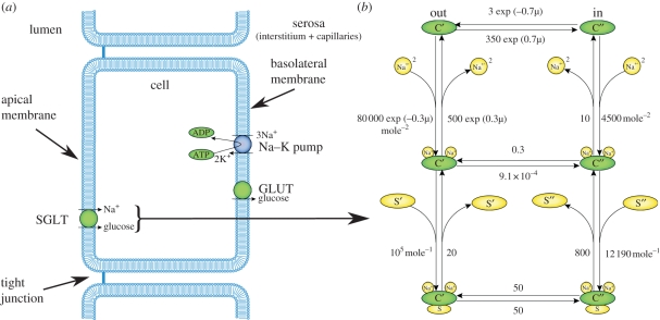
Figure 1.
(a) Schematic diagram illustrating the structure of an epithelial cell of a renal tubule including three of the transport proteins found in the proximal tubule (PT), the sodium–glucose cotransporter (SGLT) located on the apical membrane and the sodium–potassium pump (Na–K pump) and facilitated glucose transporter (GLUT) located on the basolateral membrane. (b) The state transition diagram for the Eskandari et al. [1] SGLT1 model, a specific instance of a model representing the generic transport mechanism shown in (a).

