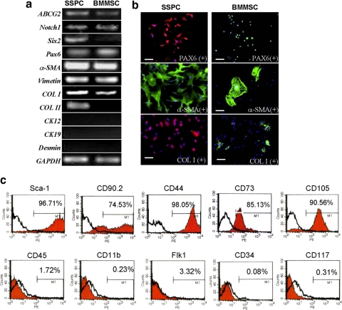
Figure 2.
Phenotype for SSPCs. (a) RT-PCR analysis of gene expression profiles related to stem cells (ABCG2 and Notch1), neural crest origin (Six2), eye development (Pax6), sclera (a-SMA, vimentin, and collagen I), and keratocytes (Ck12, Ck19, and desmin). (b) Immunostaining for proteins related to scleral tissue and stem cells in mouse SSPCs and BMMSCs. Bars, 100 μm. (c) Flow cytometry analysis for the expression of cell surface markers related to mesenchymal stem cells (Sca-1, CD90.2, CD44, CD73, and CD105), hematopoietic stem cells (CD34, CD117), leukocytes (CD45), macrophages (CD11b), and endothelial cells (Flk-1) on mouse SSPCs.

