Figure 1.
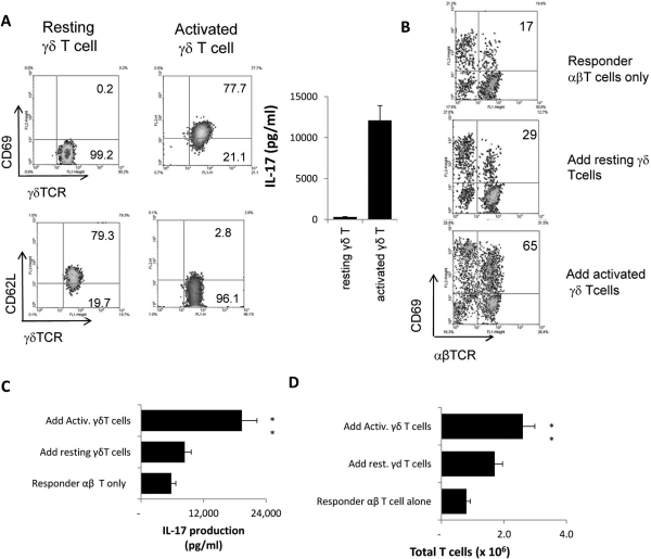
Activated γδ T cells promote αβ T cell response. (A) Phenotypes of resting and activated γδ T cells, showing that activated γδ T cells expressed increased levels of CD69 and decreased levels of CD62L. Activated, but not resting, γδ T cells produced significant amounts of IL-17. (B) In vivo–primed IRBP-specific responder αβ T cells expressed increased levels of CD69 during in vitro stimulation in the presence of activated, but not nonactivated, γδ T cells. Responder αβ T cells (1 × 10⁶/well) from immunized TCR-δ−/− mice were subjected to antigenic stimulation for 2 days by exposure to immunizing antigen and APCs, with or without the addition of 2% (2 × 10⁴/well) of activated or resting γδ T cells. Then activated T cells were separated on Ficoll and stained for the expression of CD69 and either αβTCR or γδTCR. The numbers indicated in the upper right quadrants are calculated percentage values of CD69+ cells among the αβTCR+ cells. (C) ELISA assay for cytokine production after 2 days of in vitro stimulation. IL-17 in the culture supernatants were assessed by ELISA. (D) Assessment of total number of T cells after 5 days of in vitro stimulation. The results shown are representative of those from multiple (>10) experiments. **P ≤ 0.01; differences were considered very significant.

