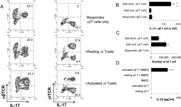
Figure 2.
Activated γδ T cells promote, whereas nonactivated γδ T cells inhibit, the activation of IRBP-specific IL-17+ αβ T cells. (A) Intracellular staining for IL-17+ αβ and γδ T cells. Responder αβ T cells (1 × 10⁶/well) prepared from IRBP1–20 immunized TCR-δ−/− mice on day 5 after immunization were stimulated for 2 days with immunizing antigen and APCs under Th17-polarized conditions, with or without the addition of 2% (2 × 10⁴/well) of resting or activated γδ T cells. Numbers indicated in the upper right quadrants are calculated percentage values of IL-17+ cells among the αβTCR+ (left) and γδTCR+ (right) cells. (B, C) Total number of IL-17+ αβ T cells and γδ T cells among the responder T cells in (A). (D) BMDCs (5 × 10⁵/well) were cocultured with resting or activated γδ T cells (1 × 10⁵) for 48 hours. Culture supernatants were tested by ELISA. The results shown are representative of those from five experiments. *P ≤ 0.05; differences were considered significant. **P ≤ 0.01; differences were considered very significant.

