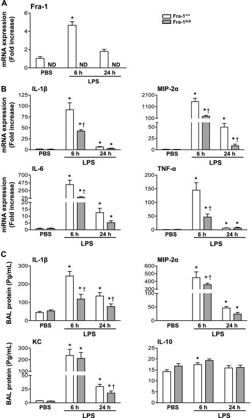
Figure 4.
Expression of inflammatory cytokines in lungs of Fra-1+/+ and Fra-1Δ/Δ mice after instillation of LPS. Fra-1+/+ and Fra-1Δ/Δ mice were instilled with PBS (n = 4 for each genotype) or LPS (10 μg/ mouse) (n = 6 for each genotype), and killed after 6 hours or 24 hours. (A) Quantitative RT-PCR analysis of Fra-1 expression in the lung after administration of LPS. (B) Quantitative RT-PCR analysis of LPS-induced inflammatory cytokines in the lung. (C) Levels of cytokines in cell-free BAL fluid, as analyzed by Bioplex assay and ELISA (MIP-2α). *P < 0.05, PBS versus LPS. †P < 0.05, Fra-1+/+ mice versus Fra-1Δ/Δ mice.

