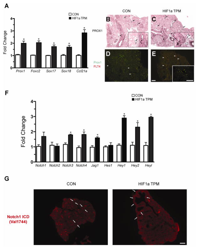
Figure 10.
Epithelial HIF1a TPM expression induces lymphangiogenesis in the embryonic lung. A) qPCR analysis of lymphangiogenic markers in HIF1a TPM lungs on dox from E6.5 until harvest at E14.5. n=3–5 samples per genotype. *p<0.05 vs. CON. B–E) PROX-1 immunostaining in E14.5 lungs. Note increased numbers of PROX1+ endothelial cells in lymphatic vessels of HIF1a TPM lungs (C vs. B, arrows) and PROX1+ staining in neuroendocrine cells of the epithelium (B and C, arrowheads). An increase in the number and size of PROX1+/FLT4+ lymphatic vessels was also observed in HIF1a TPM lungs compared to CON (panels D and E, arrowheads). Scale bars = 100μm. F) qPCR analyses demonstrates increased expression of the Notch signaling pathway genes in HIF1a TPM lungs. n=3–6 samples per genotype. *p<0.05 vs. CON. G) Notch1 signaling is increased in the mesenchyme subtending the proximal epithelium of HIF1a TPM lungs (arrows), as demonstrated by immunofluorescent staining with the cleaved Notch1 antibody Val1744. Scale bars = 200μm.

