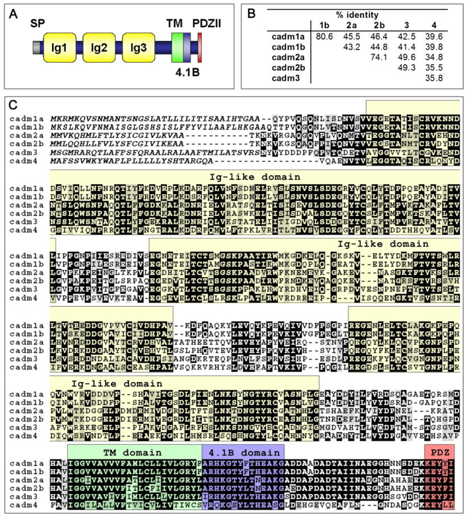
Fig. 1.
Structure and sequence of zebrafish Cadm proteins. A: Schematic representation of Cadm protein structure. All Cadms contain a signal peptide (SP), three Ig-like domains (Ig), a transmembrane (TM) domain, and a cytoplasmic tail with a 4.1B binding domain (4.1B) and the PDZ type II binding domain (PDZII). B: Amino acid identity as a percentage in pairwise alignments for the six Cadm protein sequences. C: Protein alignment of the zebrafish Cadm protein sequences. Conserved positions with an identical amino acid (black) and conserved subtitutions (gray) have been shadowed. Putative signal peptide for each cadm is in italic; each structural domain is indicated above the aligned sequences and color shaded as in A.

