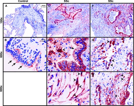
Figure 1.
Nuclear β-catenin staining is observed in systemic sclerosis (SSc)-associated pulmonary fibrosis. (A) Low (100×) and (B) high (600×) magnification images from a control lung show β-catenin staining at the cell–cell contacts of airway epithelial cells (arrows) but no obvious staining in nuclei of alveolar epithelial or interstitial cells. Control lungs are taken from normal donors whose lungs were not used for transplant. (C–H) Lung section images from patients with SSc-associated pulmonary fibrosis show β-catenin staining at junctions of airway epithelial cells (D, arrows), in nuclei of alveolar epithelial cells (G, arrowheads), and within cytoplasm and nuclei of cells that appear to be fibroblasts (E and H, arrowheads). Images are representative findings from three control patients and three patients with SSc–interstitial lung disease using Zeiss Axioskop with a CRi Nuance multispectral camera.

