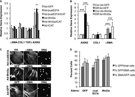
Figure 3.
Wnt/β-catenin signaling is not sufficient to induce expression of TGF-β or its established profibrotic targets in NL-57 and WI-38 human fibroblasts. (A) Lung fibroblasts (NL-57) obtained from a patient without lung disease were treated with Ad-GFP (control), Ad-S37A–β-catenin (20 PFU/cell), Ad-Wnt3a (10 PFU/cell), or Ad-ICAT (20 PFU/cell) for 24 hours; total RNA was extracted to quantify expression of α-SMA, COL1, TGF-β, and AXIN2 mRNAs by quantitative PCR. Results are expressed as the mean gene expression ± SD relative to GAPDH from three independent experiments (*P < 0.05; **P < 0.01). (B) Human embryonic fibroblasts (WI-38) were infected with Ad-GFP (control) or Ad-Wnt3a (10 PFU/cell) after undergoing treatment with control or differentiation media for 3 days; total RNA was extracted to quantify expression of AXIN2, COL1, and α-SMA mRNAs by qPCR. Results are expressed as the mean gene expression ± SD relative to GAPDH from three independent experiments (***P < 0.001). (C) Immunofluorescence of NHLFs infected with Ad-GFP, Ad-Wnt3a-GFP, and Ad-S37A–β-catenin–HA for 24 hours. Immunostaining was performed to confirm the presence of hemagglutinin (HA), GFP, and α-SMA; cell nuclei were stained with Hoechst. (D) Infection efficiency with Ad-GFP, Ad-S37A–β-catenin, and Ad-Wnt3a is routinely > 40% and expressed as GFP- or HA-positive cells divided by the total number of cells (%GFP/total cells). More than 50% of α-SMA–expressing cells are infected, expressed as GFP- or HA-positive cells divided by number of α-SMA–expressing cells (%GFP/SMA). Similarly, more than 50% of infected cells are α-SMA positive (%SMA/GFP). Results are expressed as the percent ± SEM from three independent experiments.

